
“There is no more difficult art to acquire than the art of observation, and for some men it is quite as difficult to record an observation in brief and plain language.” – Sir William Osler, MD, CM
With the first year of medical school under my belt, I’ve received the expected types of questions from family and friends: “Are you happy where you are?”, “Did you like it?”, “Was it what you expected?”, and “Will you take a look at this?” The answer to all of the above being “yes”, “yes”, “yes”, and “please see an actual medical professional.” My favorite question, however, is “what was your favorite class?”
The Art of Observation
It is often said that medicine is both an art and a science, a statement that took on a deeper significance during my selective, the Art of Observation. This course offered a chance to explore the oft-overlooked humanities component of our medical education by focusing on the critical appraisal of artistic work and examining the collaborative and intertwined history of both art and medicine.
Our first exercise was to do something most medical students are reluctant to do: tell everyone why he or she has the right answer in front of the doctor. After being shown different paintings of various scenes, we were asked to interpret what was shown to us – the time of day, period of history, mood of the scene, etc. We were then asked why we thought what we thought, forced to defend our hypotheses with visual evidence in the work – shadows cast by the sun, the fashion of the dresses worn, the expressions on people’s faces, so on and so forth. Further still, we were asked to objectively examine what led us to our conclusions based on fact alone. We supposed that the scene was taking place in the morning because of sunlight breaking through a window. But what else is sunlight other than a decrease in the value of colors over a certain area? Is a window not four rectangles circumscribed by a larger rectangle? And do we know it is made of glass because colors and shapes can be seen within the rectangle that are different from the colors and shapes outside the rectangle?
Our second exercise focused on visuospatial assessments and interpretation of 3D images. Chop a sphere in half and what do you get? A circle of course. And a cube? Obviously, a square. What about a cone? Depending on the angle of the bisecting plane a circle, ellipse, and triangle could be the right answer. Maybe I should have paid more attention in geometry and calculus. You utilize these skills to interpret an MRI or mentally reconstruct a 3D image (or even physically reconstruct a 3D image) based on 2D data. It’s also an important skill in understanding the 2D limitations of tests like radiography. This exercise was followed-up with a charcoal drawing studio session with a nude model and more painting analyses in the gallery. During this class, we practiced bringing our observations and interpretations to life. After being told to draw and erase a few times and meditating on the idea that nothing is permanent, I put together a piece of work that I’m actually very proud of (as a beginner of course).
Medicine, like art, requires us to sift through sand to find gold. A skill learned in this course, such as tolerance for ambiguity, is important because the right answer is rarely ever as obvious as we would like it to be. Often times, we are forced to with sort through mountains of ambiguous information in search of patterns that lead us to the correct diagnosis or treatment. One might even say that tolerance for ambiguity allows us to appreciate and validate different perspectives, a skill that is useful when consulting your colleagues or trying to have a conversation with a patient about his or her fears or goals. It is useful for us to tolerate ambiguity and uncertainty as a step in the learning process, instead of perceiving it as a threat or an obstacle. Other skills learned in this course, such as spatial awareness, might even be useful for assessing facial expressions and body language in your patients and colleagues, a necessary facet of emotional intelligence.
Another challenge, as Sir Osler stated, is “record[ing] an observation in brief and plain language.” Trying to explain a disease process to a layperson is like trying to explain to someone what their reflection is if they’ve never seen a mirror before. It’s an exercise that one can easily write off as trivial because we know what a mirror is from previous experience. But when you’re explaining to a patient why you think a their proximal muscle weakness is polymyositis versus a limb girdle muscular dystrophy for example, your own previous experience is going to mean little to them if you can’t explain why using simple terms to parse difficult concepts into digestible information.
This course really was my favorite class, but it was something that I didn’t fully appreciate until we finished our musculoskeletal-dermatology-rheumatology block. “Having a patient walk can tell you a lot about what’s wrong with them”, “a rash isn’t just a rash”, and “we don’t really know what’s going on here” are a few of the important points I picked up. A few weeks ago, I spent a Saturday at the MoMA, excited that I knew why Paul Klee’s painting style differed from one painting to another (it was systemic sclerosis, of course). Do me a favor the next time you’re at a museum: tilt your head or crouch down on the floor. There could be a whole world in front of you that you never even knew existed, if you only look a little harder.
I’m going to close this post with a little interactive session. Below are a few images that were used during my course, each of which depicts a certain medical condition or inconsistency (highlight under the picture to reveal the answer or discussion – best viewed on desktop). Make an observation, defend your hypothesis with evidence in the painting, and have fun.
– TS℞
The Anatomy Lesson of Dr. Nicolaes Tulp, Rembrandt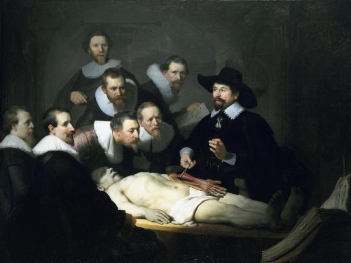
- The proportions of the body are off. The head is off-center to the right and looks propped up or devoid of a neck. It also looks a little larger than what would be expected for the body. The right arm is shorter than the left and does not reach past his hip. These inconsistencies may be due to multiple iterations of the painting using various cadavers with varying proportions. Click for article.
Rubens and the Graces, Rubens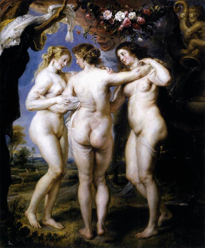
- One of the Graces might have arthritis. The leftmost figure displays a Boutonnière deformity on the fingers of her right hand (the distal interphalangeal joints are flexed while the proximal interphalangeal joints are extended).
The Ugly Duchess, Massys
- The Duchess may have had Paget’s disease, a disorder of bone deposition in which unchecked osteoclast activity leads to accelerated and disorganized deposition of lamellar bone.
The Death of Procris (A Satyr mourning over a Nymph), di Cosimo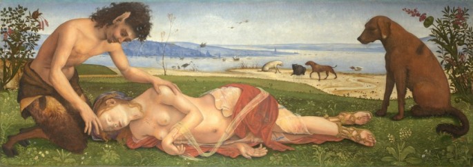
- The adducted, medially rotated, extended arm and pronated forearm would lead me to believe that the nymph developed a condition called Erb’s palsy/waiter’s tip due to a lesion of her C5-C6 nerve roots (which makes sense given the location of her injury – the cricoid cartilage). One might also look at lacerations on her forearm and posit that they were sustained while defending herself against the satyr (who are known in mythology for their voracious sexual appetites). I don’t know, I’m not a forensic pathologist.

Love your blog. Lifestyle posts pertaining to medical students is such a fresh breath of air contrary to the usual preachy study tips fr med students kind of blogs.
Ps- This one’s my favorite post 🙂
LikeLike
Thank you so much! Never really thought my writing would reach anyone so this was awesome to read 🙂
LikeLike